Key Takeaways
- AI-assisted radiology can provide faster preliminary findings, but image analysis and complete clinical interpretation are distinct capabilities.
- SignalPET combines automated radiographic analysis, context-aware reporting, PACS, and access to board-certified veterinary radiologists within one platform.
- Veterinary radiology AI platforms differ in anatomical coverage, reporting depth, specialist access, and integration with existing imaging workflows.
- Automated findings should be evaluated alongside patient history, image quality, clinical presentation, and the consequences of a missed or incorrect finding.
- Veterinary practices should assess how AI fits into their existing diagnostic process, including escalation, documentation, turnaround time, and professional oversight.
Veterinary radiology is entering a new phase in which artificial intelligence is becoming part of routine diagnostic workflows rather than a separate technology used only for selected cases. General practitioners increasingly have access to automated image analysis, structured reporting, and specialist consultation without sending every radiographic study through a conventional referral process.
The clinical challenge, however, extends beyond identifying abnormalities on an X-ray. Veterinarians need to interpret imaging findings alongside patient history, presenting symptoms, and the decisions that must be made during a consultation. A technically accurate observation may have limited clinical value if it arrives too late, lacks sufficient context, or does not help determine the appropriate next step.
Top 5 AI Tools for Veterinary Radiology
1. SignalPET
SignalPET is an AI-powered veterinary radiology platform that combines automated image interpretation, clinically contextualized reporting, imaging infrastructure, and specialist support. Its integrated approach addresses a recurring limitation of standalone AI screening tools: identifying a radiographic abnormality is not always sufficient to help a veterinarian determine what that finding means for an individual patient. SignalPET offers different reporting pathways according to the level of interpretation required.
Its Immediate Report provides rapid AI screening, while the Complete Report incorporates information such as patient history, signalment, clinical presentation, and the veterinarian’s specific questions. This allows the system to connect radiographic observations with the broader clinical picture rather than interpreting images entirely in isolation.
Key features
- Immediate AI-powered radiographic screening
- Context-aware Complete Reports incorporating patient history
- Automatic escalation to board-certified radiologists
- Traditional signed radiologist reports
- Integrated web-based PACS
- Structured diagnostic findings and clinical recommendations
- Support for standardized workflows across veterinary groups
2. Vetology
Vetology combines automated radiographic analysis with veterinary teleradiology services, allowing practices to use AI for initial interpretation while retaining access to specialist reporting. Its AI system analyzes canine and feline radiographs and generates structured findings across supported anatomical regions.
This provides a preliminary assessment that veterinarians can incorporate into their clinical evaluation, particularly when they need timely information during routine appointments or emergency consultations. The platform’s combination of automated analysis and conventional teleradiology allows practices to distinguish between cases that benefit from rapid screening and those requiring additional specialist interpretation.
Key features
- AI-assisted analysis of canine and feline radiographs
- Automated structured findings and reporting
- Thoracic, abdominal, and musculoskeletal screening
- Access to veterinary teleradiology services
- Specialist reporting for cases requiring additional interpretation
- Digital image submission and reporting workflows
3. Antech RapidRead
Antech RapidRead is an AI-assisted veterinary radiology solution associated with Antech’s broader diagnostic services. Its role is to provide automated radiographic interpretation that can help veterinarians obtain timely information during patient assessment. This is relevant to general practices where imaging is frequently performed during consultations and clinicians need to determine whether additional testing, monitoring, treatment, or specialist involvement may be appropriate.
RapidRead belongs to the category of tools that emphasize automated analysis and reporting, rather than functioning primarily as a conventional teleradiology referral service.
Key features
- AI-assisted veterinary radiographic interpretation
- Automated imaging reports
- Rapid diagnostic support for clinical workflows
- Digital radiology capabilities
- Integration considerations within Antech’s diagnostic ecosystem
- Support for veterinarian-led assessment and follow-up
4. Picoxia
Picoxia provides AI-assisted radiographic interpretation for veterinarians, with an emphasis on straightforward image submission and automated reporting. Its platform accepts common image formats, including DICOM, JPEG, and PNG, and provides a viewer through which clinicians can examine uploaded studies. The system supports automated analysis of thoracic, abdominal, and pelvic radiographs, including detection of supported imaging patterns and generation of written reports.
The platform’s defined anatomical coverage should be considered during evaluation, particularly by practices that perform a broad range of imaging studies. A clinic primarily interested in thoracic and abdominal radiographs may have different requirements from a referral hospital handling advanced imaging or complex multisystem cases.
Key features
- AI analysis of supported veterinary radiographs
- Thoracic, abdominal, and pelvic imaging support
- Automated written reports
- Differential diagnosis suggestions
- Confidence information for supported findings
- DICOM, JPEG, and PNG compatibility
- Integrated image viewer
5. Radimal
Radimal is a veterinary imaging service that combines AI-assisted radiographic analysis with access to veterinary diagnostic expertise. It represents a hybrid approach in which automated interpretation contributes to the diagnostic workflow while specialist consultation remains available when additional assessment is needed.
When considering Radimal, practices should distinguish the automated imaging capabilities from its specialist consultation services and confirm how those services are accessed within the current offering. The value of a hybrid platform depends partly on how easily clinicians can move from preliminary findings to more extensive review without duplicating administrative work.
Key features
- AI-assisted veterinary radiographic analysis
- Digital imaging workflows
- Access to specialist consultation
- Diagnostic reporting support
- Automated and human-assisted interpretation pathways
- Support for clinical imaging review
AI Screening and Clinical Interpretation Are Not the Same Thing
One of the most important distinctions in veterinary radiology software is the difference between detecting radiographic findings and interpreting an entire clinical case.
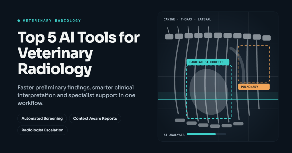
An AI system may identify an abnormal pulmonary pattern, an enlarged cardiac silhouette, or a suspected skeletal abnormality. These observations can help direct the veterinarian’s attention and provide an additional layer of screening.
Clinical interpretation requires more context.
Consider two dogs with similar thoracic radiographic findings. One presents with an acute respiratory complaint, while the other has undergone imaging as part of an unrelated investigation. Their histories, examination findings, and clinical circumstances may lead to different differential diagnoses and follow-up decisions.
This is why the information included in an AI report matters as much as the speed at which it is generated.
A platform that detects abnormalities can provide useful screening assistance. A system that incorporates clinical history and the veterinarian’s diagnostic question can provide a different level of support. Neither removes the veterinarian’s responsibility to interpret findings within the complete clinical picture.
For veterinary practices, the appropriate reporting model depends on how imaging is used. Routine screening, emergency assessment, complex diagnostic investigations, and specialist referrals may require different levels of interpretation.
Integrating AI Into the Veterinary Radiology Workflow
Introducing AI radiology software should not require veterinarians to redesign every step of an established imaging process.
The implementation should begin with how radiographs are acquired, reviewed, documented, and discussed with clients.
An integrated AI-assisted radiology workflow
- Image acquisition and quality review
The veterinary team obtains the necessary radiographic views and checks whether the study is technically adequate. - Automated analysis
The AI system processes supported images and produces preliminary findings or a structured report. - Clinical interpretation
The veterinarian evaluates the findings alongside the patient’s history, examination, symptoms, and diagnostic question. - Specialist escalation
Complex, uncertain, or consequential cases receive additional review when appropriate. - Documentation and clinical action
The findings and their interpretation inform the medical record, client communication, and subsequent clinical decisions.
Integration with existing imaging equipment and practice management systems can reduce administrative work. However, technical integration is only one part of implementation.
Veterinary teams also need clear expectations about when AI reports can inform routine decisions, when additional investigation is necessary, and when a radiologist’s interpretation should be obtained.
For multi-location veterinary groups, standardized reporting and escalation procedures can help establish a more consistent approach across hospitals.
What Veterinary Practices Should Examine Before Implementation
A useful evaluation should focus on representative clinical cases rather than relying exclusively on demonstrations of obvious abnormalities.
Practices should include routine studies, technically challenging images, cases with multiple findings, and situations in which the imaging results may significantly influence treatment.
The evaluation should address four areas: supported anatomical regions and species, reporting depth, specialist availability, and integration with existing clinical workflows.
Diagnostic performance also requires careful interpretation. Accuracy figures are only meaningful when their underlying population, study design, conditions, and performance measures are understood. Sensitivity for one radiographic finding does not establish equivalent performance across every condition or patient population.
Operational performance deserves similar scrutiny. Fast automated reports have practical value when they arrive at the point of care and provide information that clinicians can use. A rapid result that requires extensive manual interpretation or creates additional administrative work may deliver less benefit than expected.
The objective is to introduce AI in a way that strengthens the existing diagnostic process, with clearly defined responsibilities for both automated systems and veterinary professionals.
FAQs
Can AI accurately interpret veterinary X-rays?
AI can identify supported radiographic patterns and provide automated findings, but performance varies by condition, species, anatomical region, image quality, and the system being used. An AI tool’s performance for one abnormality should not be generalized to every imaging study. Veterinarians should review relevant validation evidence and interpret automated results alongside clinical history, examination findings, and other diagnostic information.
Can AI replace a veterinary radiologist?
AI can support screening and interpretation, but it does not eliminate the need for veterinary radiologists. Specialist expertise remains important for complex findings, uncertain results, advanced imaging, and cases where imaging has significant implications for treatment. Some AI radiology platforms integrate automated analysis with access to board-certified radiologists, allowing practices to use different levels of diagnostic support according to the clinical situation.
How quickly can veterinary AI radiology software generate reports?
Turnaround times vary by platform, report type, and the complexity of the requested interpretation. Automated screening may return findings within minutes, while comprehensive AI reports and conventional radiologist interpretations can require additional time. Practices should distinguish between the availability of preliminary automated findings and the delivery of a complete diagnostic report when evaluating advertised turnaround times.
What should veterinary clinics look for in AI radiology software?
Veterinary clinics should assess supported species and anatomical regions, diagnostic validation, report quality, turnaround time, image format compatibility, and integration with existing systems. Access to specialist consultation may also be important. Testing the software with representative clinical cases helps establish whether its capabilities align with the practice’s imaging volume, diagnostic requirements, and established clinical workflows.
Is AI veterinary radiology useful for emergency hospitals?
AI-assisted radiology can provide timely preliminary findings during emergency assessment, particularly when immediate access to a radiologist is limited. Its usefulness depends on supported imaging studies, diagnostic performance, and how results are incorporated into clinical decisions. Emergency hospitals should maintain appropriate escalation procedures because automated screening cannot reliably exclude every serious condition or replace specialist interpretation when it is clinically indicated.